Editor’s Summary: The essay “Anatomy of the Human Cardiovascular System” explores the intricate structure and function of the human cardiovascular system. It details the roles of the heart, blood vessels, and blood in maintaining homeostasis by delivering oxygen and nutrients while removing waste. The heart is highlighted as the central pump with four chambers that circulate blood, while the transport network of arteries, veins, and capillaries facilitate blood flow. The composition and functions of blood, including red and white blood cells and platelets, are also discussed. The essay underscores the importance of coronary circulation and regulatory mechanisms in ensuring cardiovascular efficiency.
Anatomy of the Human Cardiovascular System Essay

The human cardiovascular system is a complex network of organs and vessels that work in harmony to circulate blood throughout the body. This intricate system plays a crucial role in maintaining homeostasis by delivering oxygen and nutrients to tissues while removing waste products. This essay will explore the anatomy of the cardiovascular system, focusing on its major components and their functions.
Heart: The Central Pump
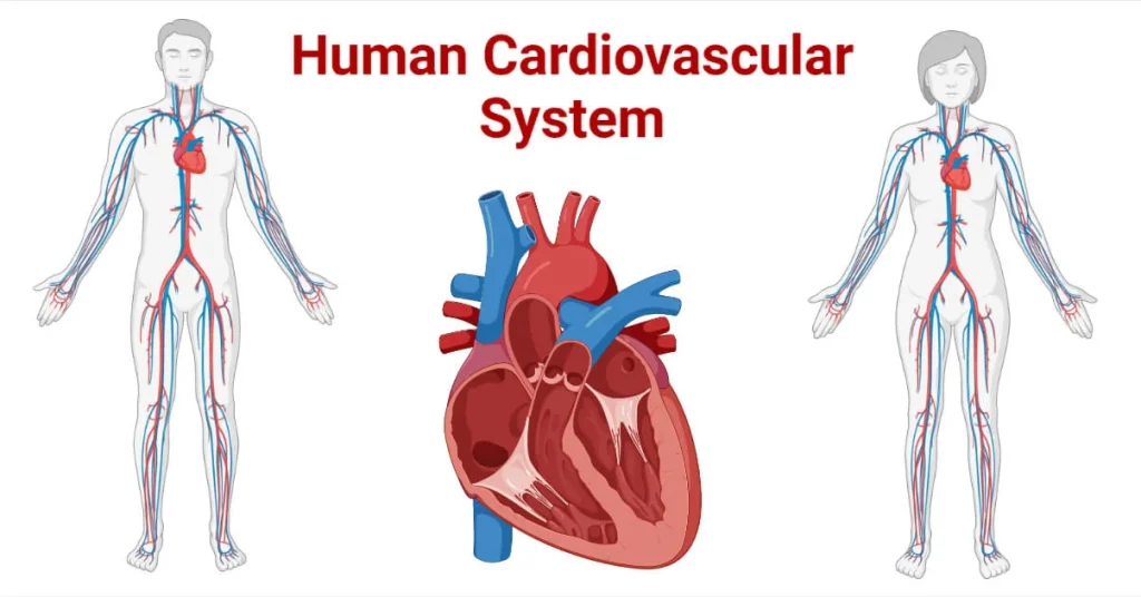
At the core of the cardiovascular system lies the heart, a muscular organ roughly the size of a closed fist. The heart is located in the thoracic cavity, slightly left of center, and is protected by the ribcage and surrounded by the pericardium, a double-layered membrane (Marieb & Hoehn, 2018). Structurally, the heart is divided into four chambers: two upper atria and two lower ventricles.
The right atrium receives deoxygenated blood from the body via the superior and inferior vena cavae. This blood then flows through the tricuspid valve into the right ventricle, which pumps it to the lungs through the pulmonary arteries. In the lungs, the blood releases carbon dioxide and picks up oxygen. Oxygenated blood returns to the left atrium via the pulmonary veins, passes through the mitral valve into the left ventricle, and is then pumped out to the body through the aorta (Tortora & Derrickson, 2017).
The heart’s ability to pump blood effectively relies on its specialized cardiac muscle tissue, the myocardium. This tissue is unique in its ability to contract rhythmically without external stimulation, thanks to the sinoatrial node, the heart’s natural pacemaker.
Blood Vessels: The Transport Network
The cardiovascular system’s transport network consists of three main types of blood vessels: arteries, veins, and capillaries. Each type has a specific structure suited to its function.
Arteries carry blood away from the heart under high pressure. They have thick, elastic walls to withstand this pressure and maintain blood flow. The largest artery, the aorta, branches into smaller arteries and then into arterioles, which regulate blood flow into capillary beds (Scanlon & Sanders, 2018).
Capillaries are the smallest blood vessels, with walls only one cell thick. This thin structure allows for efficient exchange of gases, nutrients, and waste products between the blood and surrounding tissues. The extensive network of capillaries ensures that nearly every cell in the body is close to a blood supply.
Veins carry blood back to the heart under low pressure. They have thinner walls than arteries but contain valves to prevent backflow of blood. Small veins, or venules, collect blood from capillary beds and merge into larger veins. The largest veins, the superior and inferior vena cavae, return deoxygenated blood to the right atrium of the heart (Marieb & Hoehn, 2018).
Blood: The Transport Medium
Blood is the fluid medium that circulates through the cardiovascular system, consisting of plasma and formed elements. Plasma, which makes up about 55% of blood volume, is a straw-colored liquid composed mainly of water, proteins, and other dissolved substances. The formed elements include red blood cells (erythrocytes), white blood cells (leukocytes), and platelets (thrombocytes).
Red blood cells, which give blood its characteristic color, are responsible for oxygen transport. They contain hemoglobin, a protein that binds to oxygen in the lungs and releases it to tissues throughout the body. White blood cells play a crucial role in the immune system, defending the body against pathogens and other foreign substances. Platelets are cell fragments essential for blood clotting, helping to prevent excessive blood loss when blood vessels are damaged (Tortora & Derrickson, 2017).
Coronary Circulation: Supplying the Heart
The heart muscle itself requires a constant supply of oxygen and nutrients to function properly. This is achieved through the coronary circulation, a specialized part of the cardiovascular system. The right and left coronary arteries, which branch off from the aorta just above the aortic valve, supply blood to the heart muscle. These arteries divide into smaller branches that penetrate the myocardium, ensuring adequate blood supply to all parts of the heart (Scanlon & Sanders, 2018).
After passing through capillaries in the heart muscle, deoxygenated blood is collected by coronary veins. Most of this blood drains into the coronary sinus, a large vein that empties into the right atrium. The coronary circulation is critical for heart health, and blockages in these vessels can lead to serious conditions such as myocardial infarction (heart attack).
Regulation of Cardiovascular Function
The cardiovascular system is regulated by both intrinsic and extrinsic mechanisms to ensure it meets the body’s changing needs. Intrinsic regulation includes the heart’s ability to adjust its output based on the volume of blood returning to it (Frank-Starling mechanism) and the autoregulation of blood flow in tissues based on local conditions.
Extrinsic regulation involves the autonomic nervous system and various hormones. The sympathetic nervous system can increase heart rate and contractility, while the parasympathetic system has the opposite effect. Hormones such as epinephrine and norepinephrine can also influence cardiovascular function, increasing heart rate and blood pressure in response to stress or exercise (American Heart Association, 2022).
Conclusion
The human cardiovascular system is a marvel of biological engineering, combining structural complexity with functional elegance. From the rhythmic contractions of the heart to the intricate network of blood vessels and the specialized functions of blood components, each part of the system plays a crucial role in maintaining the body’s internal environment. Understanding the anatomy of the cardiovascular system is essential for medical professionals and forms the foundation for diagnosing and treating a wide range of cardiovascular disorders.
References:
1. Marieb, E. N., & Hoehn, K. (2018). Human Anatomy & Physiology (11th ed.). Pearson.
2. Tortora, G. J., & Derrickson, B. (2017). Principles of Anatomy and Physiology (15th ed.). Wiley.
3. Scanlon, V. C., & Sanders, T. (2018). Essentials of Anatomy and Physiology (8th ed.). F.A. Davis Company.
4. American Heart Association. (2022). How the Healthy Heart Works. Retrieved from https://www.heart.org/en/health-topics/congenital-heart-defects/about-congenital-heart-defects/how-the-healthy-heart-works
